
Gel + bioceramics
In order to apply robust hydrogels for clinical applications of next generation artificial cartilage, we developed a method to enable atoxic fixation of gels to bone tissue. Strong adhesion at the gel-bone interface has been demonstrated in animal experiments.
1. The bonding of soft tissues to hard tissues in nature
There are many living things that possess supporting tissues in nature. Supporting tissues play an essential role in the body, and include both hard tissues (bone) and soft tissues (tendon, ligament, skin, fascia etc.). In contrast to tissues involved in body homeostasis such as organs and blood cells, these tissues play an important role in the body by providing fixation and allowing vigorous exercise and smooth motion. Hard and soft tissues are intricately connected to enable these functions in living things. If you have ever eaten fried chicken or chicken wings, you are probably aware that bones are very strongly connected to tendons and cartilage. How are these two tissues connected? Organisms have achieved the robust connection by forming an uninterrupted continuous structure at the boundary between the two tissues that have completely different compositions and physical properties.
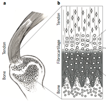
At the boundary, there is a gradual transition from soft to hard tissue. This will act as a guideline for developing new bonding materials. Contrasting these structures for fixing biomaterials in vivo, currently, medical string and medical glue have been used, but both are unsatisfactory for hydrogels that contain a lot of water, like cartilage tissue. Therefore, the development of a new bonding method to fix water-rich materials is required.
2. Creation of osteoconductive hydrogel (HAp/DN gel)
Biomaterials currently used as artificial soft tissues for medical use are hard and dry materials such as metals, ceramics, and plastics. These materials have much harder physical properties than actual living soft tissues (such as cartilage, ligaments, and tendons). This can cause issues in the body, such as causing damage to the hard tissues to which they are attached. Therefore, it is necessary to switch to soft and wet materials that more closely match the physical properties of soft tissue. For example, double network (DN) gels developed by our laboratory possess high strength and toughness and exhibit excellent compressive mechanical properties, comparable to that of living cartilage. It has been expected to find application as a next-generation artificial cartilage material. In addition, DN gel is the first material known to induce regeneration of cartilage tissue in vivo, so it is expected to also be a useful material for the reconstruction of cartilage tissue.Soft tissues are always connected with other tissues (e.g. bone) in vivo. However, as mentioned above, it is difficult to bond gel materials to bones. We solved this problem by mimicking the biological mechanism. Bone is a natural organic-inorganic composite composed of organic collagen (20%) and inorganic hydroxyapatite (HAp) (80%), and is a tissue with excellent strength and toughness. Bones are capable of naturally repairing damage when broken. Taking advantage of this property, a HAp coating is applied to artificial bones and dental implants prior to use. In our laboratory, we have extended this technique to soft tissues, by developed an osteoconductive HAp / DN gel that can be firmly bound to bone.
As you can see from the movie, even though fragile HAp is incorporated, the HAp / DN hybrid gel is very tough and flexible, and HAp is chemically stable in the gel for long times because the HAp nanoparticles are physically interlocked with the gel network on the nano-scale.
3. Elucidation of the bonding mechanism of HAp/DN gel to bone by animal experiments
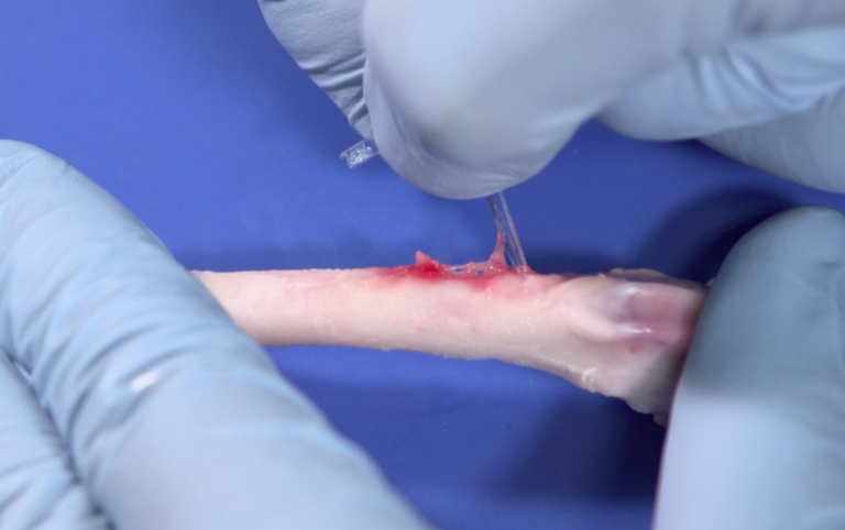
We have demonstrated via animal experiments that this HAp / DN gel can be firmly adhered to bone in vivo.
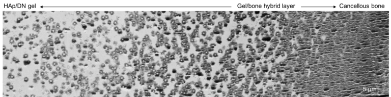
The videos are the results of a mechanical test one month after implanting HAp / DN gel into the skull and femur of a rabbit, respectively. The gels are firmly bonded to both parts, and the adhesive strength exceeded the breaking strength of the DN gel. To observe the details of the structure achieving this high adhesive strength, we used a transmission electron microscope to examine the bone-gel interface structure. As shown in the photo, it is confirmed that the boundary between hydrogel and bone did not show a clear border; rather a gradient structure is clearly visible.
This structure is very similar to the boundary between bone and cartilage (ligament, tendon) in natural tissue, and it should be an ideal adhesion method for living organisms. At present, we are not limited to just basic research. We are working on controlling the local adhesion site to bone for three dimensional complicated surfaces, and also evaluating the effect on cells by performing cell culture studies on HAp / DN gels.
References
- Genin, G. M. & Thomopoulos, S. Unification through disarray.Nat. Mater. 16, 607–608 (2017).
- Gong, J., Katsuyama, Y., Kurokawa, T. & Osada, Y. Double-Network Hydrogels with Extremely High Mechanical Strength.Adv. Mater. 15, 1155–1158 (2003).
- Yasuda, K. et al. A Novel Double-Network Hydrogel Induces Spontaneous Articular Cartilage Regeneration in vivo in a Large Osteochondral Defect.Macromol. Biosci. 9, 307–316 (2009).
- Kiyama, R. et al. Micro patterning of hydroxyapatite by soft lithography on hydrogels for selective osteoconduction.Acta Biomater. 81, 60–69 (2018).
- Nonoyama, T., Wada, S., Kiyama, R. & Kitamura, N. Double-Network Hydrogels Strongly Bondable to Bones by Spontaneous Osteogenesis Penetration.Adv. Mater. 28, 6740–6745 (2016).
- Wada, S., Kitamura, N., Nonoyama, T., Kiyama, R. & Kurokawa, T. Hydroxyapatite-coated double network hydrogel directly bondable to the bone : Biological and biomechanical evaluations of the bonding property in an osteochondral defect. Acta Biomater. 44, 125–134 (2016).